Fluorescence TR-MOKE System
Merging Time-Resolved Magnetization and Fluorescence Detection
Discover the advanced time-resolved MOKE (TR-MOKE) and fluorescence system that combines time-resolved magnetization with fluorescence detection technologies such as NV-Centers and Excitons. This innovative approach allows researchers to achieve unprecedented insights into magnetic materials and their dynamics. With our state-of-the-art tabletop system, you can easily observe and analyze magnetic phenomena with high spatial and temporal resolution. Ideal for both academic and industrial applications, the Fluorescence TR-MOKE system opens new frontiers in materials science and condensed matter physics.
Specifications
- Fluorescence and TR-MOKE Microscopy at the Same Time
- Individual Excitation and Detection Spectra
- Spatial Resolution < 350 nm
- Temporal Resolution < 500 fs
- Magnetic Field Range > 500 mT
- Magnetic Field Angle: up to 360 °
- Individual RF Sources
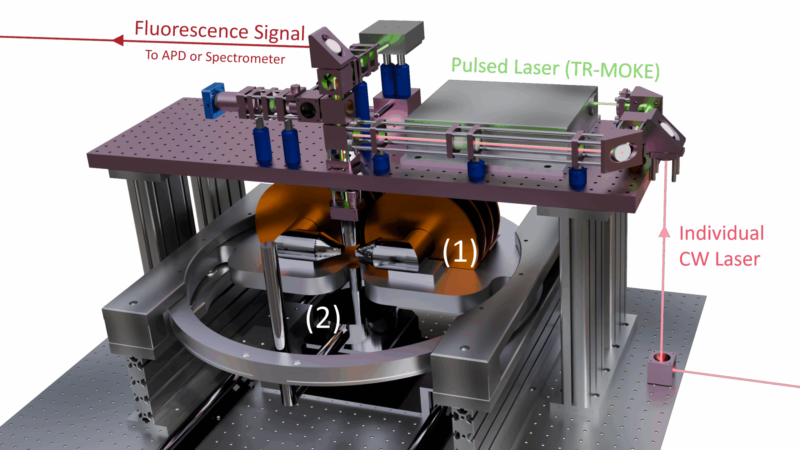
- Individual Magnetic Fields (1)
- Low Temperature Option (2)
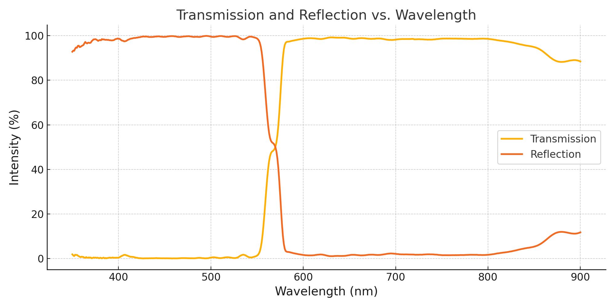
Example Spectra for Excitation and Detection optics
Our Fluorescence TR-MOKE system allows for full customization of excitation wavelengths and detection spectral ranges, enabling targeted interaction with a wide variety of fluorescent centers or materials. Whether your research focuses on defect centers, quantum emitters, or general photoluminescent phenomena, we can configure the optical path accordingly.
The standard configuration is optimized for NV center microscopy, supporting simultaneous time-resolved MOKE and fluorescence detection. This dual-mode capability allows for correlated studies of magnetic and optical properties, crucial for cutting-edge research in quantum sensing, spin dynamics, and nanoscale magnetometry.
Get in touch with us to discuss a custom spectrum configuration tailored to your experiment.

Laser -Sources
The choice of laser wavelength and repetition rate plays a key role in time-resolved magneto-optical Kerr effect (TR-MOKE) experiments, directly impacting spatial resolution, signal sensitivity, and the accessible time window. To meet a wide range of experimental demands, we offer a flexible selection of femtosecond and picosecond laser sources.
For lower GHz-range dynamics, picosecond lasers provide a cost-effective alternative with sufficient temporal resolution. In fluorescence microscopy applications, especially those involving NV centers, we also provide continuous-wave (CW) laser sources tailored to specific excitation needs.
Our standard femtosecond systems include 800 nm (400 nm SHG) and 1040 nm (520 nm SHG) lasers, with repetition rates from 1 MHz to 100 MHz and pulse energies up to 100 nJ—custom configurations available on request.
- Standard Lasers:
- 1040 nm and 520 nm (via SHG)
- 800 nm and 400 nm (via SHG)
- Repetition rates: 1 MHz – 100 MHz
- Pulse energy: up to 100 nJ (higher energies on request)
Get in touch with us to discuss a custom laser configuration tailored to your experimental setup.
rf -SourceS
Our proposed RF systems offer high-frequency precision up to 67 GHz, enabling tailored excitation of magnetic systems across a broad spectrum—from low-GHz ferromagnetic resonance (FMR) measurements to high-frequency spin dynamics.
Fine-tuning the RF frequency is critical for selectively addressing specific magnetization modes and dynamic regimes. This precision not only supports TR-MOKE applications but also enables targeted excitation of NV centers, allowing for controlled spin manipulation and advanced quantum sensing experiments.
Whether your research focuses on dynamic magnetic responses or quantum spin control, the RF sources used for our Fluorescence TR-MOKE are built for accuracy and stability.
Get in touch with us to discuss your specific needs.
Electromagnet options
Our systems feature robust electromagnets engineered for high-precision magnetization experiments. With in-plane magnetic fields exceeding 1 Tesla, they deliver the stability and strength required for dynamic magnetic studies. Out-of-plane configurations are also available upon request to accommodate specialized experimental needs.
For full flexibility, our setups support up to 360° rotation of the in-plane magnetic field, enabling angular-dependent measurements without sample repositioning—essential for anisotropy characterization. An integrated water-cooling system ensures thermal stability, even under sustained high-field operation.
To match various experimental demands, we propose both unipolar and bipolar power supplies, providing precise control over field polarity and amplitude.
Get in touch with us to discuss your specific needs.
explore TR-MOKE
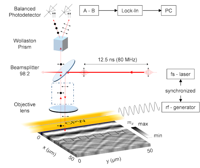
Schematic overview of a standard TR-MOKE setup. Femtosecond laser
pulses are used for stroboscopic measurement of coherently excited spin waves. A galvo-galvo unit can be used for scanning the laser pulses across the sample surface (placed at
the position marked by the dashed rectangle). Alternatively, the sample can be moved
via a xyz piezo scanning stage to scan the sample surface with respect to the laser focus.
Both systems are offered by CRI²SPIN.https://nbn-resolving.org/urn:nbn:de:bvb:91-diss-20230324-1693644-1-1
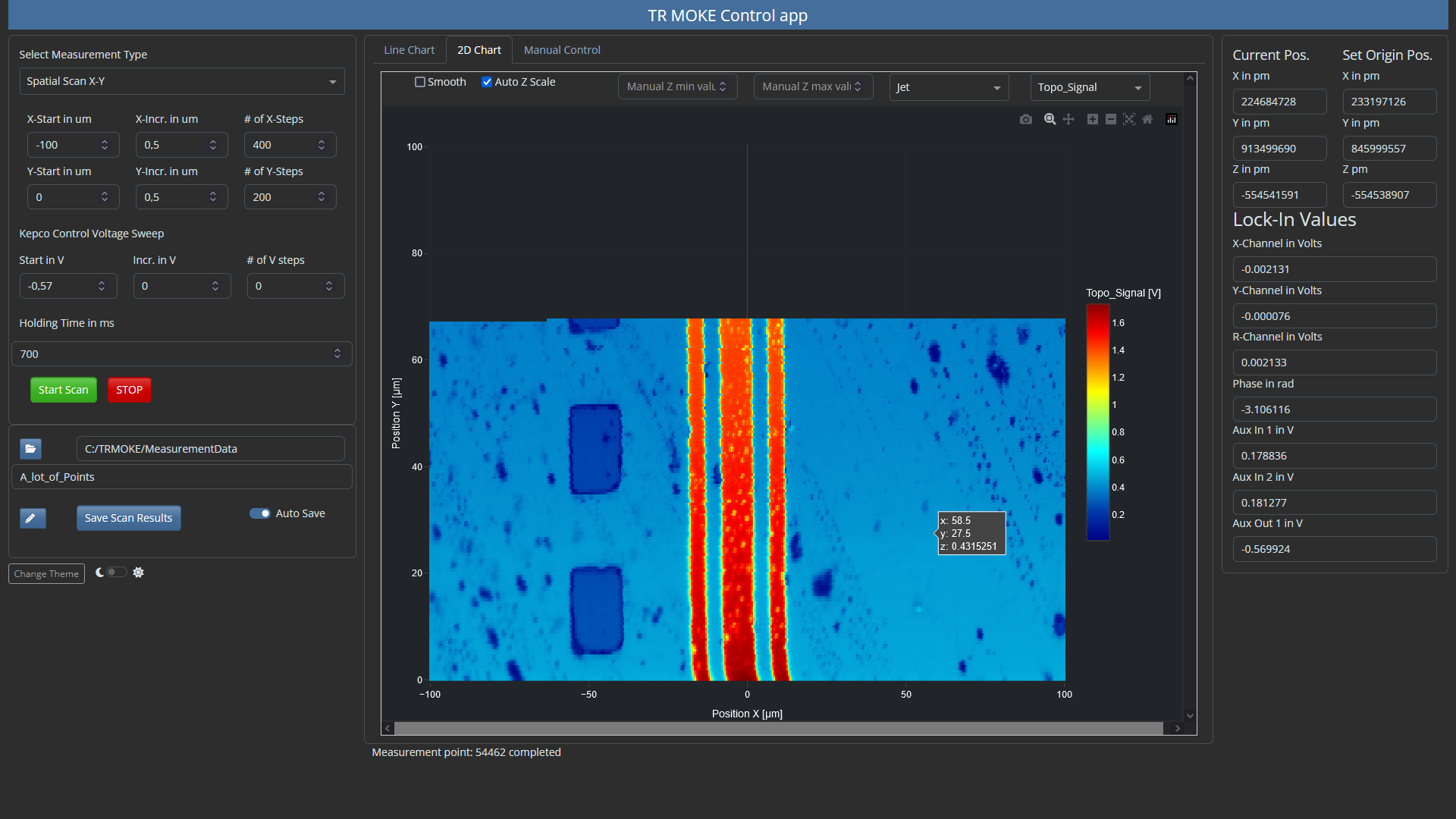
TR-MOKE user software with example data for topographic signal (measurement in progress). Automatic result report pdf file can be created to achieve “Labbook-ready” documentation.
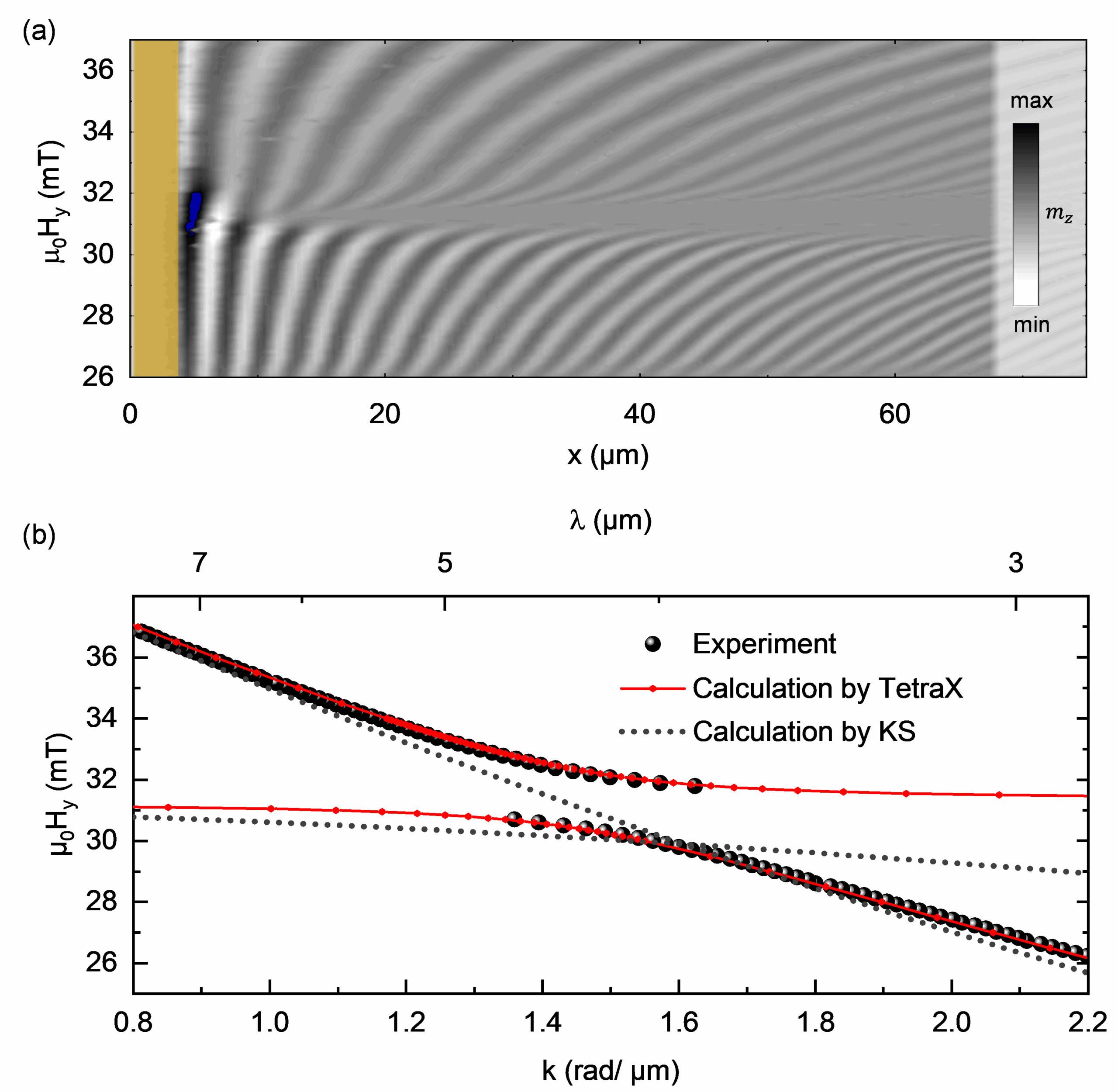
(a) Spin wave propagation as a function of the external magnetic field. An area with high attenuation is observed around 31 mT.
(b) Black spheres show the extracted k-values from TR-MOKE measurements presented in (a). The dispersion relation calculated using TetraX (red symbol-line) is in good agreement with the experimental data, whereas the calculation using the zeroth order perturbation theory from Kalinikos and Slavin (KS) does not give the proper solution in the vicinity of the hybridization.
https://nbn-resolving.org/urn:nbn:de:bvb:91-diss-20230324-1693644-1-1
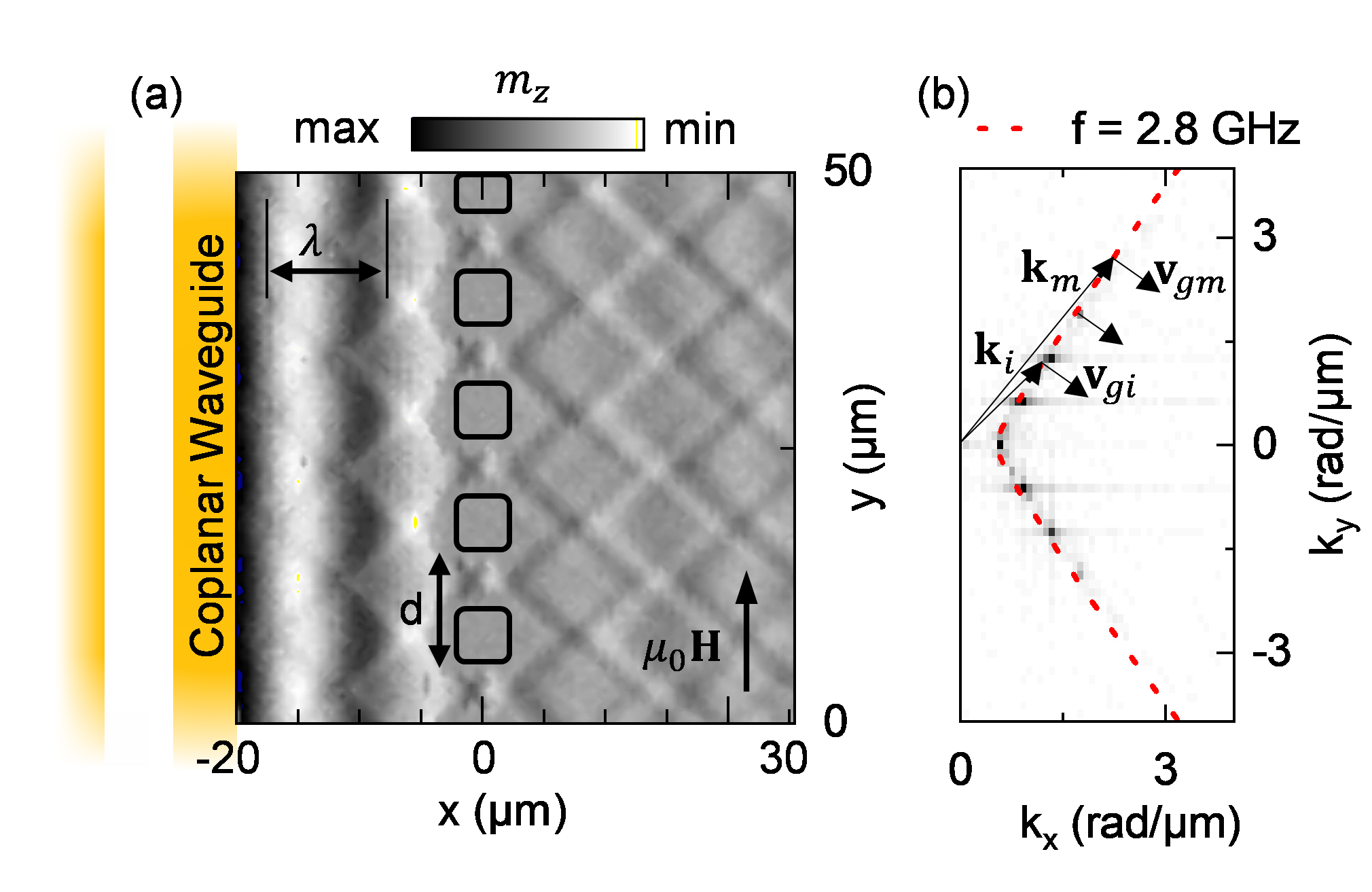
TR-MOKE measurement of a plane spin wave propagating
through a diffraction grating, indicated by the black squares. (a) Caustic-like spin wave beams are emitted from a point like source at the diffraction grating in both, the positive and negative x-direction. (b) Corresponding 2D FFT of the region behind the diffraction grating (x > 0) representing the emitted wave-vectors in both, x- and y-direction.C. Riedel et al., (2023), Adv. Phys. Res., 2: 2200104
Let’s talk
Tell us your specific needs!